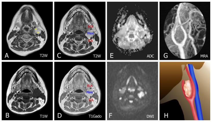
Figure 4.
42-year-old female with a left carotid body tumor.
FINDINGS: (A, C) Axial T2-weighted images show a lesion with increased signal and well-defined flow voids (yellow arrows in A). (B, D) Pre- and post-contrast T1-weighted show a mass centered in the left carotid bifurcation with avid enhancement (E, F) DWI shows evidence of restricted diffusion along with the ADC map. (G) Enhanced MR angiography maximum-intensity projection image shows tumoral enhancement and proximal splaying of the internal (ICA) and external carotid artery (ECA). (H) Illustration demonstrating the carotid body tumor in this case.
TECHNIQUE: Siemens Magnetom Amira MRI scanner, magnetic strength = 1.5 Tesla, intravenous contrast was administered.
A: Axial T2W. TR = 4000 ms. TE = 110 ms. Slice thickness = 4 mm.
B: Axial T1W pre-contrast. TR 600ms. TE 15ms. Slice thickness = 4 mm.
C: Axial T2W. TR = 4000 ms. TE = 110 ms. Slice thickness = 4 mm.
D: Axial T1W post-contrast with IV contrast in venous phase, Gadovist 15mL. TR 670 ms. TE 10ms. Slice thickness = 4 mm.
E: Axial ADC. TR = 3500 ms. TE = 90 ms. Slice thickness = 4 mm
F: Axial DWI. TR = 3500 ms. TE = 90 ms. Slice thickness = 4 mm
G: Maximum intensity projection (MIP) of a contrast enhanced magnetic resonance angiography (CE-MRA)
H: Illustration

