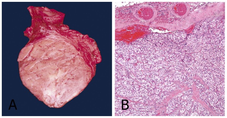
Figure 5.
42-year-old female with a left carotid body tumor.
FINDINGS: (A) The operative specimen of the carotid body tumor. It is dark-purple in color and fairly well circumscribed. (B) Hematoxylin and eosin (H&E) stain showing a well-developed Zellballen (“cell balls” in German) growth pattern. The neoplastic cells demonstrate an eosinophilic granular cytoplasm and round hyperchromatic nuclei.
TECHNIQUE:
A: Operative specimen.
B: Light microscopy, magnification 10x, hematoxylin-eosin stain.

