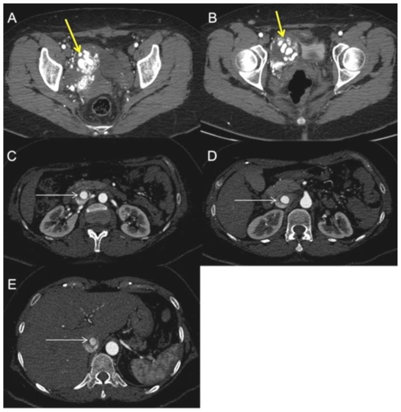
Figure 3.
A 48-year-old woman with uterine intravenous leiomyomatosis and intracardiac extension.
Technique: Contrast enhanced CT, 249 mA, 120kV, 2.5 mm slice thickness, intravenous contrast: 130 mL of Ultravist (Iopromide). GE LightSpeed 64.
Findings: A, B) Axial contrast enhanced CT of the abdomen in the arterial phase demonstrated the presence of a voluminous pelvic mass with significant contrast enhancement due to hypervascularization (see the yellow arrow). C, D, E) Notice the presence of contrast-enhanced large vessels within the IVC (see the thin white arrow).

