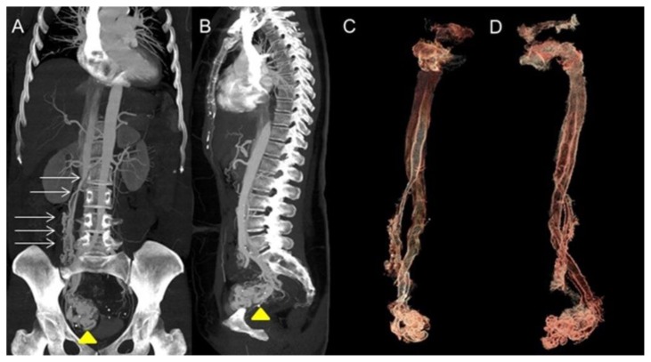
Figure 4.
A 48-year-old woman with uterine intravenous leiomyomatosis and intracardiac extension.
Technique: Contrast enhanced CT, 249mA, 120kV, 2.5 mm slice thickness, intravenous contrast: 130 mL of Ultravist (Iopromide). GE LightSpeed 64.
Findings: A,B) Coronal and sagittal CT Maximum Intensity Projections (MIP) of the abdomen in the arterial phase showing the pelvic tumor (see the yellow arrowhead) directly extending into the IVC (see the thin white arrows).
C,D) 3-D reconstruction from the source data confirmed the origin of the thrombus from a uterine mass invading the right iliac vein and then the IVC.

