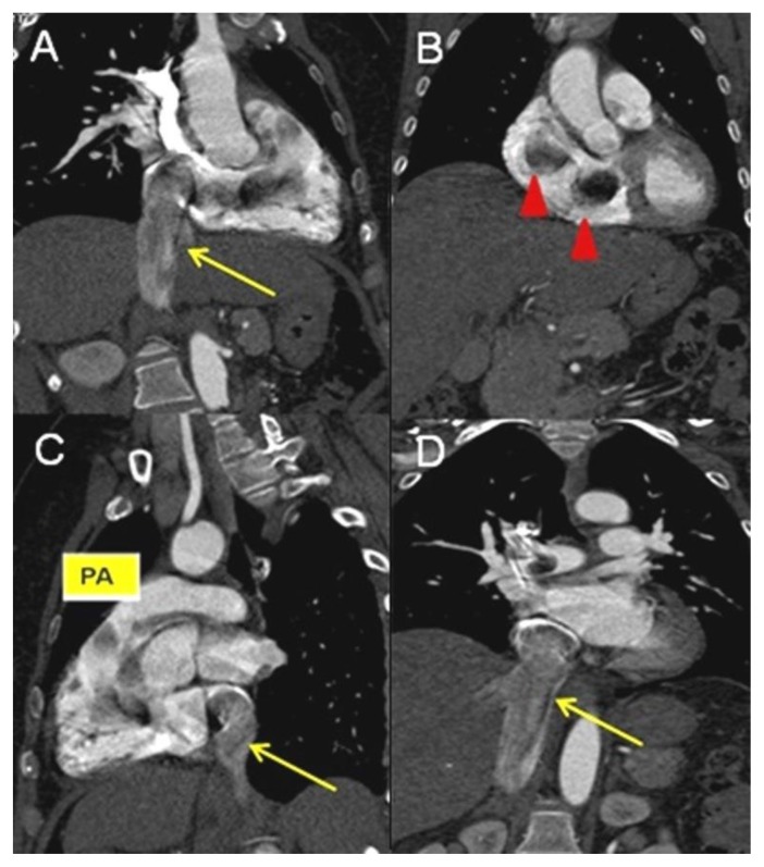
Figure 5.
A 48-year-old woman with uterine intravenous leiomyomatosis and intracardiac extension.
Technique: Contrast enhanced CT, 249mA, 120 kV, 2.5 mm slice thickness, intravenous contrast: 130 mL of Ultravist (Iopromide). GE LightSpeed 64.
Findings: A, B, C, D) Coronal and sagittal CT projections showing the caval (see the yellow arrow) and cardiac extension of the tumor, up to the right atrium, right ventricle (see the red arrowheads), right ventricle outflow, to the pulmonary trunk (pa).

