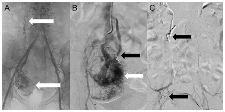
Figure 7.
A 48-year-old woman with uterine intravenous leiomyomatosis and intracardiac extension.
Technique: Selective embolization of the uterine mass performed using microcatheter Direxion (Direxion HI-FLO™ Torqueable Microcatheters Boston Scientific) and metallic coils.
Findings: A, B) The angiogram showed a hypervascular pelvic mass, with anomalous peripheral new vessels formation, invading the IVC (see the white and black arrows). C) A final angiogram showed a drastic reduction of the vascularization (see the black arrows).

