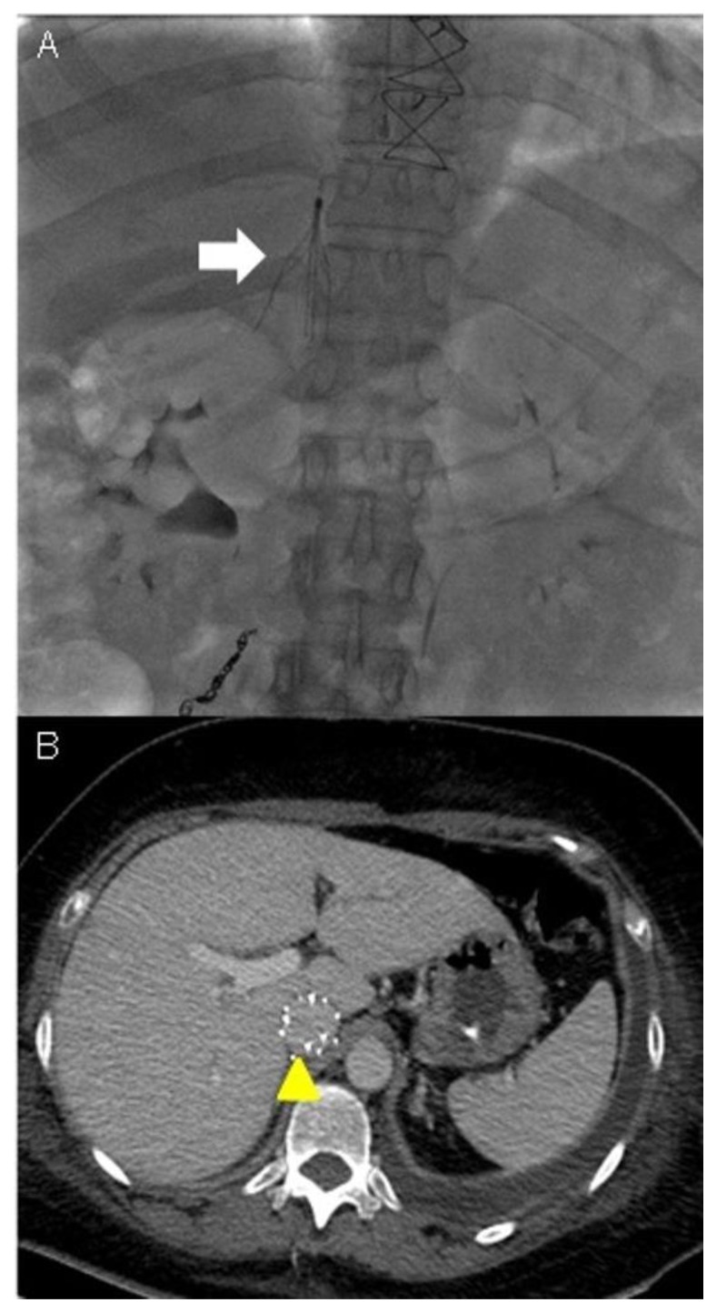
Figure 9.
A 48-year-old woman with uterine intravenous leiomyomatosis and intracardiac extension.
Technique: Under radiologic guidance a catheter was inserted via jugular vein access and advanced to the inferior vena cava in the abdomen. Contrast material was then injected into the vein to assess for proper positioning of the IVC filter.
Findings: A) The filter was correctly expanded and attached itself to the walls of the IVC (see the white arrow). B) CTA was used to monitor the results of IVC caval filter. CTA venous phase showed the caval filter in the retrohepatic IVC, of note the anomalous position of the filter was necessary due to the extension of the thrombosis above the renal veins (see the yellow arrowhead).

