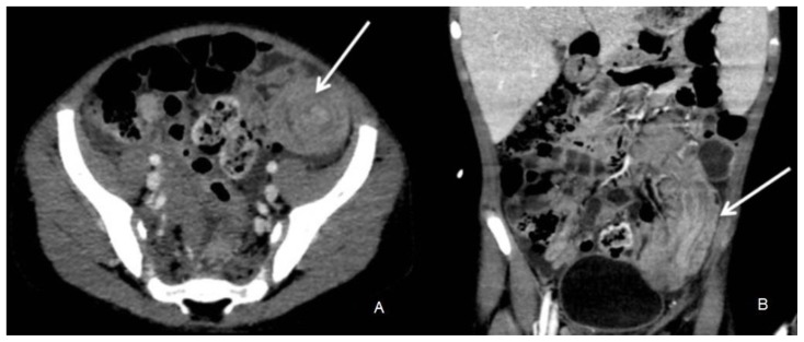
Figure 1.
6 year old boy being evaluated for Burkitt lymphoma was found to have a distal ileal intussusception.
Findings: CT demonstrates an intussusception in axial and coronal reformatted images (arrow) with invagination of proximal bowel into distal bowel loops.
Technique: Axial contrast enhanced CT abdomen and pelvis in venous phase with coronal reformats. 120 KV, 180 mAs, 1.5 mm slice thickness, 10 ml optiray 300 IV contrast.

