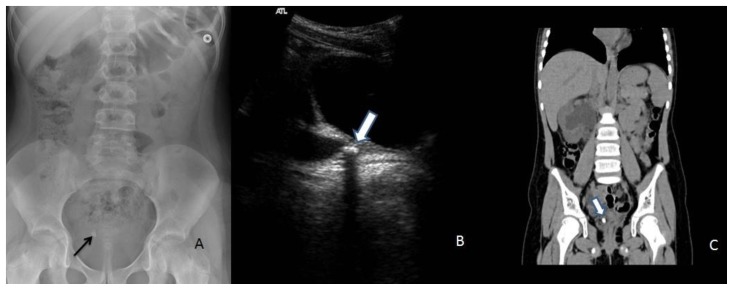
Figure 12.
9 year old girl with ureteral stone.
Findings: Abdominal radiograph (A) demonstrates a right pelvic stone (arrow).
The following two images are for the sister of the girl presented in (A). Ultrasound (B) demonstrating an echogenic focus with posterior shadowing (arrow) in the distal right ureter, with an associated dilated proximal ureter (arrowhead). On the non-enhanced CT (C) this corresponds to the calcific density in the distal ureter (arrow).
Technique: (A) Supine abdomen 80 KV, 120 mAs. (B) Real time ultrasound images of the pelvis using 5 mHz curvilinear probe. (C) Axial non-enhanced CT abdomen and pelvis with coronal reformats. 120 KV, 180 mAs, 1.5 mm slice thickness.

