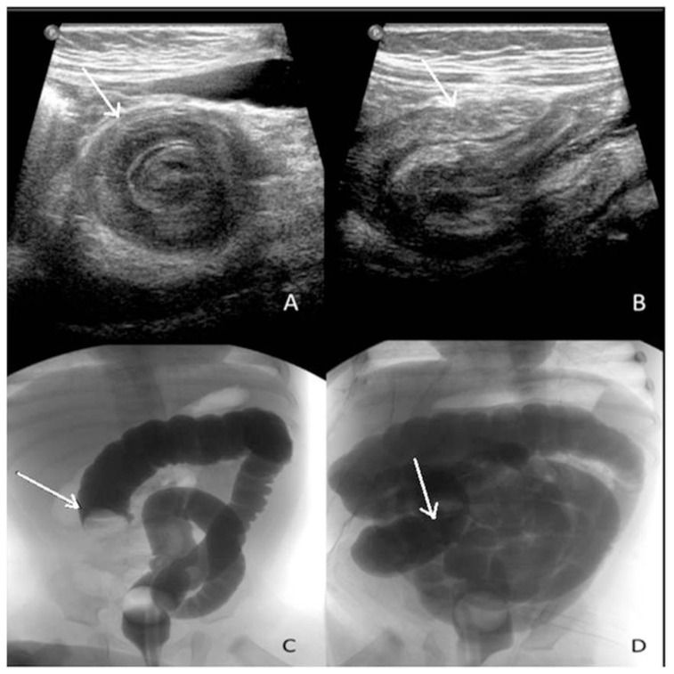
Figure 2.
5 month old boy presented to the emergency department with colicky abdominal pain, vomiting and diarrhea, found to have intussusception.
Findings: (A) Ultrasound demonstrates a target sign with a hypoechoic ring and a hyperechoic center on transverse view. (B) On the longitudinal US view, a pseudokidney sign with superimposed hypo- and hyperechoic areas representing the edematous walls of the intussusceptum and layers of compressed mucosa is observed. After confirming the lack of free air on the scout abdominal view (not shown), water soluble contrast enema (C) demonstrates a mass prolapsing in the lumen visualized as a convex shaped filling defect (arrow) that progressively reduced until contrast refluxed into the small bowels (D) with the arrow pointing to the cecum and ileocecal valve region.
Technique: (A+B) Real time ultrasound images of the right lower quadrant using 12 mHz linear probe. (C+D) Foley catheter placed in the rectum with inflation of the balloon, then, pulsed fluoroscopy following the instillation of water soluble gastrografin contrast solution.

