
Figure 3.
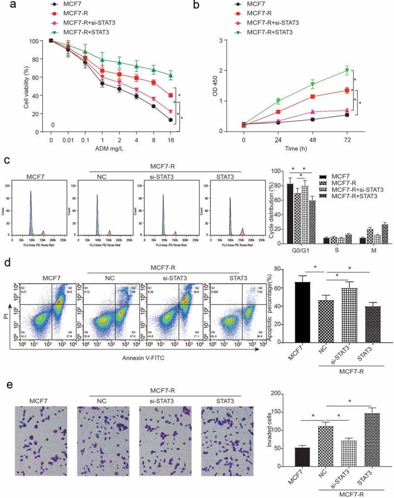
The flow apoptosis assays and cell invasion of breast cancer stem cells. (a) The cell viability of all groups decreased when the concentration of DOX increased. Compared with the MCF7-R group, the knockdown of STAT3 inhibited cell viability while the overexpression of STAT3 could promote cell viability (*P < 0.05). (b) An MTT assay was used to test the OD value at different concentrations of doxorubicin (*P < 0.05). (c) The results of the cell cycle analysis at different concentrations of doxorubicin showed that compared with the MCF-R group, the knockdown of STAT3 impeded the cell cycle in the si-STAT3 group, whereas the overexpression of STAT3 arrested the cell cycle in the STAT3 group (*P < 0.05). (d) The percentage of apoptotic cells was determined by flow cytometric analysis. The apoptosis rate was calculated by counting the percentage of early apoptotic and late apoptotic cells. The NC group had a greater apoptotic rate than the MCF7 group. Compared with the NC group, the si-STAT3 group had a greater rate of apoptosis, while the STAT3 group had a reverse consequence at both concentrations of doxorubicin. (*P < 0.05). (e) Compared with the MCF7 group, the invasion of the MCF7-R group decreased in the si-STAT3 group, whereas the invasion increased with the transfection of STAT3 (*P < 0.05).