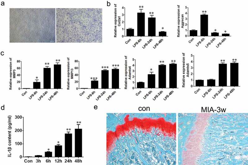
Figure 1.

Establishment of osteoarthritis models using chondrocyte cells and mice.
(a) Toluidine blue staining of chondrocytes (×100, scale bars = 1000μm). (b) Relative mRNA levels of Col2a1 and Aggrecan in chondrocytes treated with LPS were detected by Q-PCR. (c) Relative mRNA levels of MMP3, MMP13, ADAMTs4, ADAMTs5 in chondrocytes treated with LPS. (d) Content of secreted IL-1β in medium was analyzed with ELISA assay. (e) The femur tissues of the mice were stained with Safranin O (×100). Data were expressed as mean ± SEM (n = 3). Values with p < 0.05 were considered to be statistically significant. *P < 0.05, **P < 0.01, ***P < 0.001 vs. control group.
