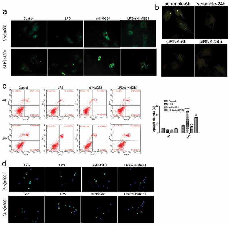
Figure 5.

HMGB1-mediated autophagic activity and apoptosis of chondrocytes.
(a) Autophagic activity was detected by MDC staining (×400). (b) Expression of intracellular GFP-LC3 was observed under the fluorescence microscope (×200). (c) Flow cytometry analysis of apoptosis in chondrocytes. (d) Detection of apoptosis in chondrocytes using TUNEL staining (×200). Data are expressed as Mean ± SEM (n = 3). Values with p < 0.05 were considered to be statistically significant. **P < 0.01, ***P < 0.001 vs. control group; #P < 0.05 vs. LPS group.
