Fig. 2.
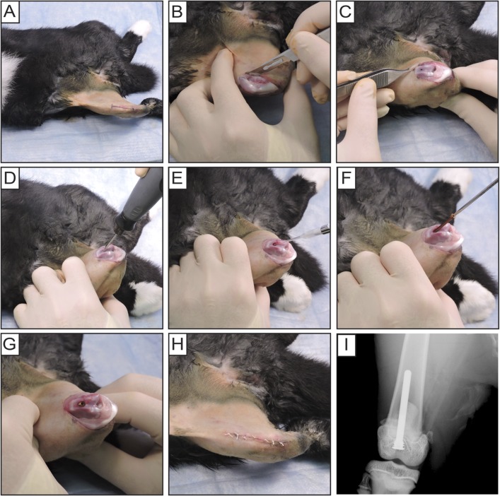
Figs. 2-A through 2-I Rabbit surgical procedures. Fig. 2-A The distal anterior region of the right thigh through the proximal aspect of the leg was shaved and prepared. Fig. 2-B After a midline incision was made over the patella, a medial parapatellar arthrotomy was performed. Fig. 2-C The patella was dislocated laterally to expose the trochlea and intercondylar notch. Fig. 2-D The femoral medullary canal was drilled and the implant was countersunk anterior to the intercondylar notch. Fig. 2-E S. aureus (bioluminescent strain SAP231, 1 × 10⁴ CFUs in 10 µL of PBS solution) was pipetted into the canal. Fig. 2-F Coated implants were manually inserted retrograde with a screwdriver. Fig. 2-G Implant flush with the articular surface. Fig. 2-H The patella was relocated and the surgical site was closed with sutures. Fig. 2-I Anteroposterior radiographic image of implant placement.

