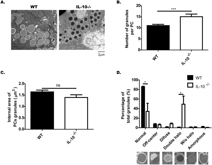
Fig 3. IL-10-/- mice present abnormal Paneth cells granules.
Paneth cell (PCs) granules of C57BL/6 and IL-10-/- mice were evaluated by transmission electron microscopy (n = 3). (A) Representative images of PC granules from both strains (4200X, Scale is 2μm). (B) Number of granules per PC (t-student, ***˂p 0.001). (C) Area of the electrondense core of the PC granules. D) Classification of granules, according to their TEM morphology in: normal, off-center, diffuse, double halo, without halo and amorphous (n = 3 mice, 2-way ANOVA, post-hoc Bonferroni *p˂0.05).

