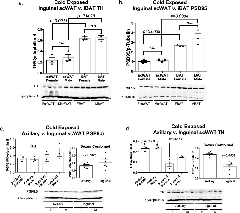
Fig 6. Adipose innervation: Sex and depot comparison.
Adult (16 week old) male and female control mice on a C57BL/6J background were cold exposed (5°C) for 3 days. Protein expression of TH (a) and PSD95 (b) were measured in inguinal scWAT and BAT via western blotting. Protein expression of pan-neuronal marker PGP9.5 (c) and sympathetic nerve marker TH (d) was measured in axillary and inguinal scWAT for both sexes via western blotting. Protein expression was normalized to β-Tubulin or Cyclophilin B, band density was quantified in Image J and analyzed using a Two-Tailed Student’s t-test. Error bars are SEMs.

