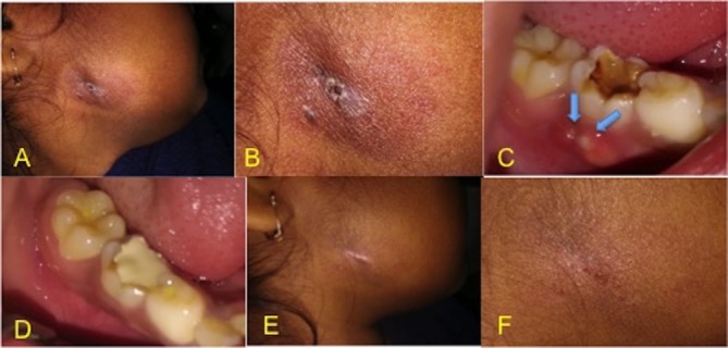
Figure 1.

(A) Extraoral cutaneous sinus tract. (B) Unhealthy granulation tissue, two stomata and erythematous surrounding skin clearly visible at the angle of jaw extraorally. (C) Tooth no. 85 showing large carious lesion and intraoral sinus tract with double stomata (arrow marked). (D) Healed intraoral sinus tract. (E) Healed extraoral sinus tract at 3 months follow-up visit. (F) Healed sinus with surrounding normal skin extraorally after 6 months, only scar mark is present.
