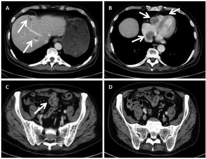
Figure 3.

Chest–abdomen–pelvis CT. (A) Hepatic metastases. (B) Low-density, non-contrasted intracavital lesion of the right atrium and ventricle in keeping with cardiac allocations of the metastatic melanoma. (C) Dotted lesions of high density in the wall of the large bowel in keeping with the metastatic lesions shown in the colonoscopy image. (D) Proximal sigmoid colon and parts of the small bowel. In contrast to the evident lesions of the large bowel, the small bowel appears unremarkable.
