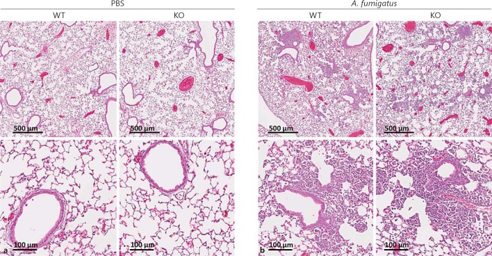
Fig. 2.
Lung histology of mice following A. fumigatus infection. WT and ficolin KO mice were intranasally inoculated with PBS as controls (a) or 2 × 10⁷A. fumigatus conidia (b) and sacrificed 24 h p.i. Lung tissues were harvested, fixed, and HE stained for histological examination. Representative lung sections from WT or ficolin KO mice are shown. Images are representative of 2 mice in PBS mock-infected groups and 6 mice in the A. fumigatus-infected groups obtained from one experiment.

