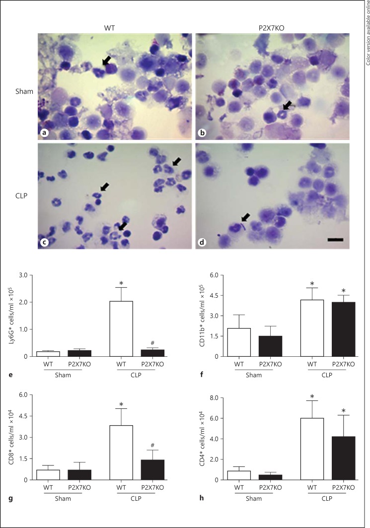
Fig. 2.
Fewer neutrophils are observed at the site of inflammation in septic P2X7KO animals. Peritoneal washes were collected 24 h after sepsis induction by CLP, and the peritoneal cell populations from Sham WT (a) Sham P2X7KO (b), CLP WT (c) and CLP P2X7KO (d) animals were analyzed by light microscopy. Arrows show neutrophils. Scale bar = 10 µm. e-h Peritoneal wash cells were also analyzed by flow cytometry using phenotypic antibody markers. Concentrations of Ly6G+ neutrophils (e), CD11b+ mononuclear cells (f), and CD8+ (g) and CD4+ (h) T lymphocytes in peritoneal washes are shown here. * p ≤ 0.05: statistically significant difference between treated and control groups (e.g. WT CLP vs. WT Sham); # p ≤ 0.05: statistically significant difference between mouse strains (e.g. WT CLP vs. P2X7KO CLP). Graphs represent results from 5 independent experiments, except for the Ly6G+ chart, which represents 3 replicates (n = 5 mice per experiment).

