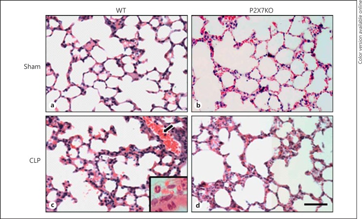
Fig. 7.
Lung tissue stained with HE portrays less damage in septic P2X7KO animals. After 24 h of sepsis induction the lungs of the animals were collected and fixed in paraffin. Cuts were made and processed for HE staining. This figure shows the lung slices of septic WT animals with an inset showing inflammatory cells (c), P2X7KO animals (d) and their respective control groups (a, b). Scale bar = 50 μm.

