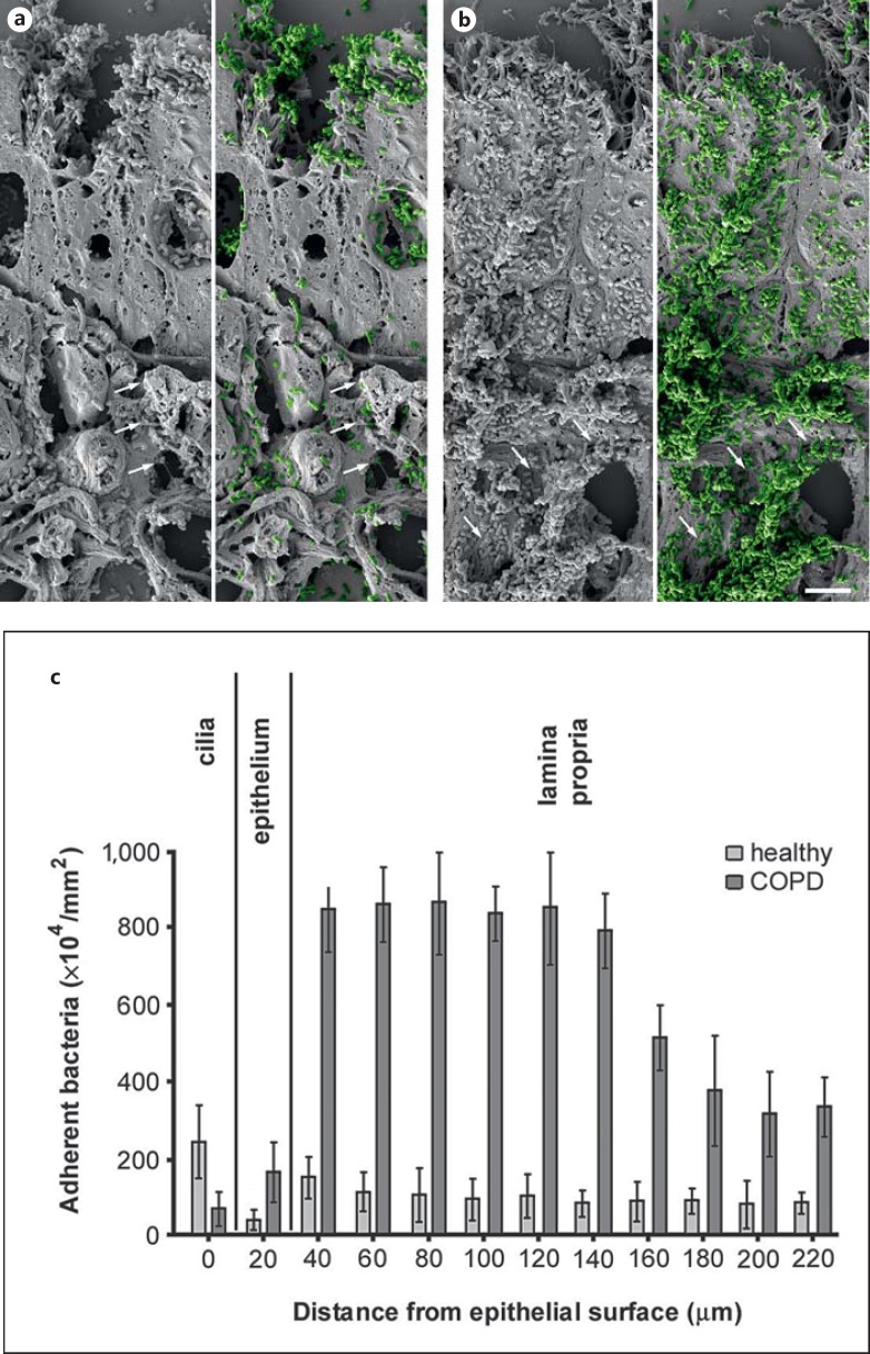
Fig. 2.
Colonization of M. catarrhalis in COPD lung tissues ex vivo. Paraffin sections of lung biopsies from healthy individuals and COPD patients were inoculated with M. catarrhalis. In COPD, large amounts of bacteria adhered to the airway mucosa (b) as compared to only moderate binding to tissues from healthy subjects (a). M. catarrhalis is highlighted in green color in a and b. Frequently bacteria were bound to fibrillar extracellular structures (arrows). The scale bar represents 5 μm. c Quantification of adherent bacteria found in different compartments of the airway mucosa.

