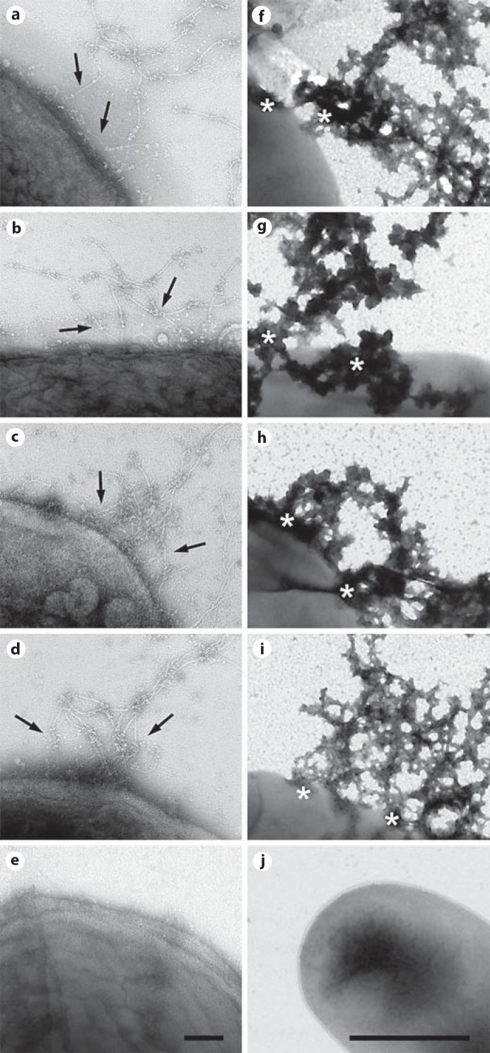
Fig. 5.
Collagen VI microfibrils adhere to M. catarrhalis and other pulmonary pathogens, followed by killing. Collagen VI microfibrils were purified from bovine cornea and allowed to react with M. catarrhalis (a), nontypeable H. influenzae (b), S. pneumoniae (c) and P. aeruginosa (d). Negative staining and transmission electron microscopy visualizes primary adhesion of collagen VI networks (arrows) to the bacterial surfaces (a-d), followed by killing within 1 h (f-i). Asterisks denote areas of membrane destabilization and exudation of intracellular content (f-i). F. magna was used as a control and did neither recruit collagen VI (e) nor exhibit killing (j). The scale bars represent 100 nm (a-e) and 1 μm (f-j).

