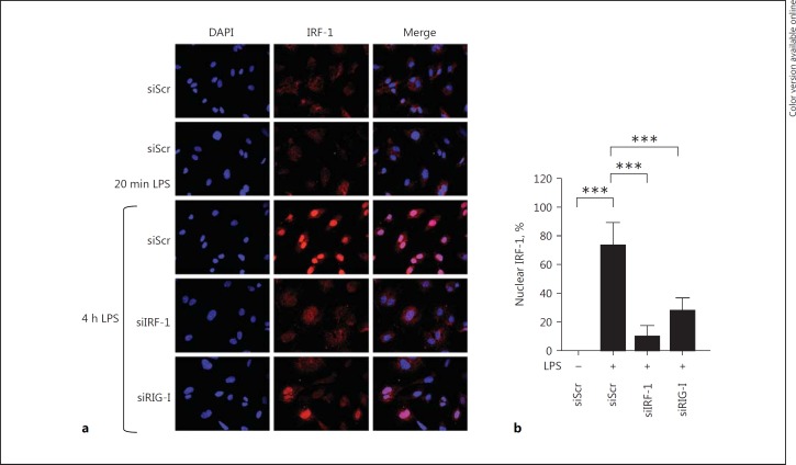
Fig. 5.
LPS-mediated nuclear translocation of IRF-1 is dependent on RIG-I. a Representative immunofluorescence images showing IRF-1 (red) and DAPI nuclear staining (blue) in HUVEC transfected with either siScr, siIRF-1, or siRIG-I and subjected to vehicle (-), 20 min or 4 h LPS. All images were taken with equal exposure times. Original magnification ×400. b The percentage of nuclear IRF-1 was quantified by analyzing at least 250 cells from each sample. Bars represent mean ± SD and data are representative of 3 independent experiments. *** p < 0.001.

