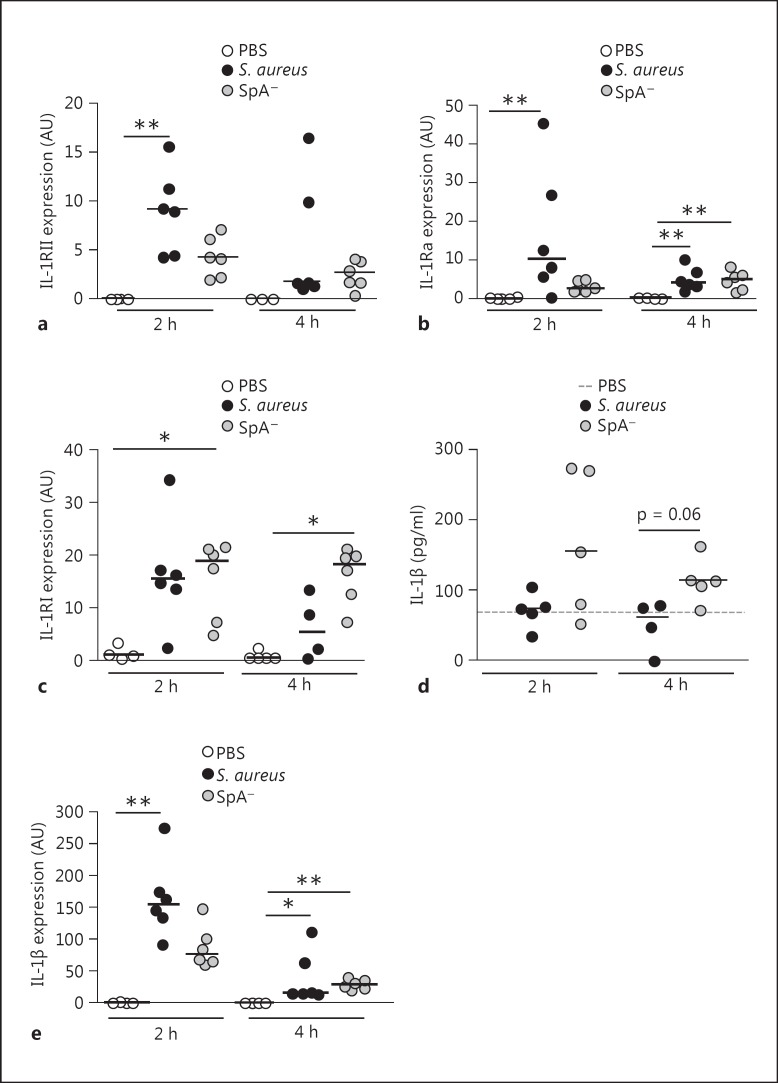
Fig. 5.
Expression of IL-1 family members during S. aureus infection. Mice were intraperitoneally inoculated with wild-type S. aureus, the SpA- mutant or PBS (control). IL-1RII (a), IL-1Ra (b) and IL-1RI (c) expression in peritoneal cells at the indicated time points after challenge was quantified by RT real-time PCR and normalized to GAPDH. Each dot represents an individual mouse and horizontal lines show the median value in each group. ** p < 0.01, Kruskal-Wallis test with Dunn's post hoc test for multiple comparisons. d IL-1β levels in peritoneal fluid were quantified by ELISA. Each dot represents an individual mouse and horizontal lines show the median value in each group (nonparametric Mann-Whitney test). e IL-1β expression in peritoneal cells at the indicated time points after challenge was quantified by RT real-time PCR and normalized to GAPDH. Each dot represents an individual mouse and horizontal lines show the median value in each group. * p < 0.05; ** p < 0.01, Kruskal-Wallis test with Dunn's post hoc test for multiple comparisons.

