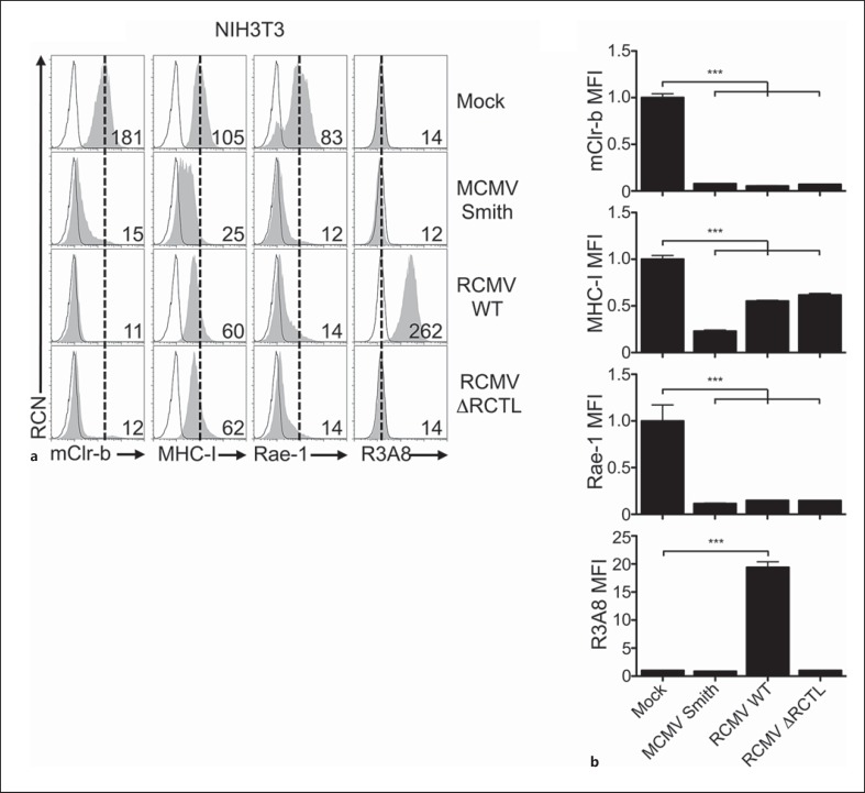
Fig. 4.
Xenogeneic RCMV-E infection promotes mouse Clr-b loss on mouse fibroblasts. a NIH3T3 were infected with MCMV-Smith or RCMV-E viruses and then analyzed by flow cytometry at 24 h.p.i. for mClr-b, MHC-I, Rae-1 and R3A8 (RCTL) expression. Primary mAb stain (shaded histograms); secondary reagent alone (black line); reference for mock-treated control MFI level (dotted vertical line). Numbers indicate MFI values. b Quantitation of MFI levels in (a) normalized to mock-treated control MFI levels. Graphs show mean ± SEM. Experiments were analyzed using one-way ANOVA with Bonferroni's post hoc analysis. * p < 0.05, ** p < 0.01, *** p < 0.001. All data are representative of at least 3 independent experiments.

