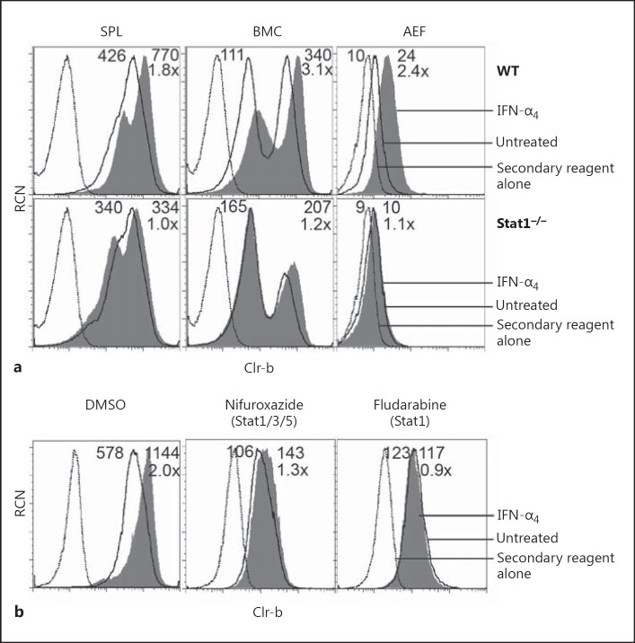
Fig. 6.
Type-I interferon (IFN)-mediated Clr-b induction is dependent on STAT1. a Splenocytes (SPL), bone marrow cells (BMC), and primary adult ear fibroblasts (AEF) were harvested from wild-type (WT) and Stat1−/− mice, treated with 103 U/mL IFN-α4 for 6 h, and analyzed by flow cytometry for Clr-b expression. b NIH3T3 cells were treated with vehicle alone (DMSO), 50 μM nifuroxazide, or 50 μM fludarabine for 22 h, at which point 103 U/mL IFN-α4 was added for 3 h prior to flow cytometric analysis of Clr-b expression. Dotted lines represent the secondary reagent alone; solid lines represent cells not treated with IFN-α4; shaded histograms represent IFN-α4-treated cells. Numbers reflect the median fluorescence intensity of untreated cells (left) or IFN-α4-treated cells (right); the lower number (e.g. 8×) represents the fold change in Clr-b MFI (IFN-α4 treated/untreated).

