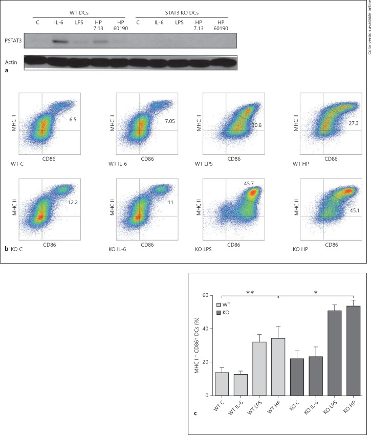
Fig. 3.
STAT3 KO DCs express enhanced levels of maturation markers in response to H. pylori infection. a Lysates from STAT3 KO (Cre+) and WT (littermate Cre− control) BMDCs incubated with H. pylori strains 7.13 and 60190 at a MOI of 25:1, IL-6 (100 ng/ml) or E. coli LPS (1 ng/ml) for 20 h in DC media were subjected to immunoblotting analysis to determine changes in STAT3 phosphorylation. b STAT3 KO and WT BMDCs were incubated with H. pylori strain 7.13 at a MOI of 25:1, IL-6 (100 ng/ml) or E. coli LPS (1 ng/ml) for 20 h in DC media. Flow cytometry analysis was used to quantify the population expressing high levels of the maturation markers CD86 and MHC class II. Cells in the gated population in the upper right quadrant of each graph represent the BMDCs with a high expression of both markers (i.e. mature DCs). c Quantification of gated CD86hi MHC IIhi DCs. Columns: means, bars: SEM. *p < 0.05; **p < 0.01 using the Kruskal-Wallis test (n = 4). a-c C = Control; HP = H. pylori strain.

