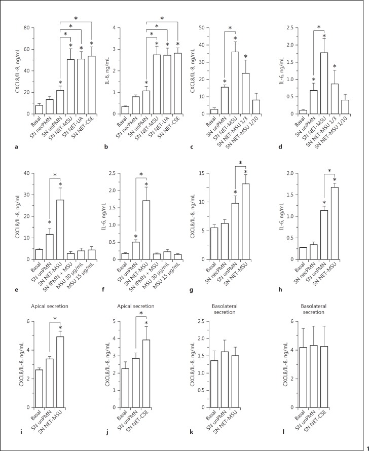
Fig. 1.
NET stimulated proinflammatory cytokine release by human epithelial cells. Cytokine secretion by A549 alveolar epithelial cells (a–f), BEAS-2B bronchial epithelial cells (g, h), or polarized 16HBE14o- human bronchial epithelial cells (i–l; apical stimulation and cytokine release to the apical [i, j] or basolateral [k, l] compartment) after 18 h of culture in the absence (Basal) or presence of supernatants of necrotic neutrophils (SN necPMN), supernatants of unstimulated neutrophils (SN unPMN), or supernatant containing NET induced by MSU (SN NET-MSU); UA (SN NET-UA) or CSE (SN NET-CSE), determined by ELISA. c, d Secretion of CXCL8/IL-8 and IL-6 by A549 cells in response to the indicated dilutions of NET released by 1 × 10⁶ neutrophils. e, f Secretion of CXCL8/IL-8 and IL-6 by A549 cells cultured in the presence of either SN NET-MSU, supernatants of PFA-fixed neutrophils (fPMN) incubated with MSU, or MSU alone. Worthy of mention, MSU was evaluated at 10- and 20-fold dilutions of that used to stimulate NETosis because, after the removal step, only a small proportion of the crystals could remain in the cultures before NETosis took place and their concentrations in NET preparations were probably lower than those tested. Data are depicted as the mean ± SEM of 7 (a), 8 (b), 3 (d, g), 4 (f, h, j, l), or 5 (e, i, k) independent experiments performed in triplicate. * p < 0.05 (two-way ANOVA with repeated measures, followed by Bonferroni's multiple-comparisons test).

