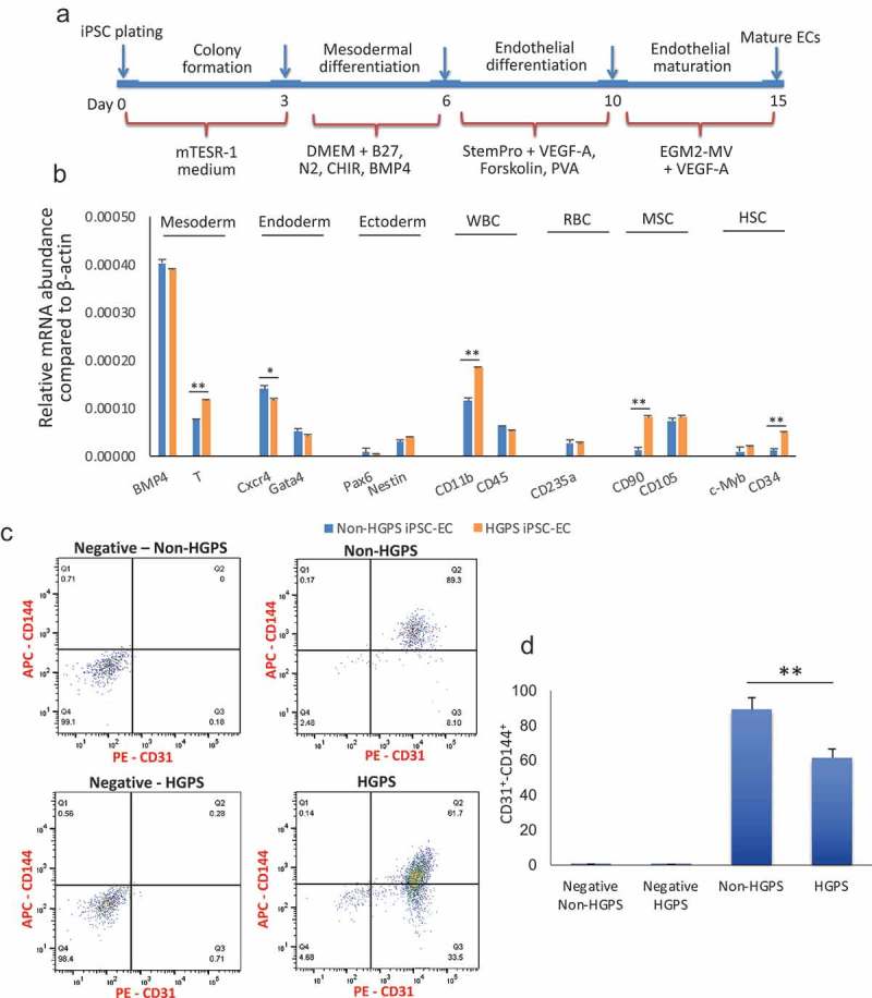
Figure 2.

Differentiation and purification of iPSCs-derived ECs. (a) Protocol for differentiation of iPSC in endothelial cells. (b) Real-time PCR for markers of mesoderm, endoderm, ectoderm, white blood cells (WBC), red blood cells (RBC), mesenchymal stem cells (MSC) and hematopoietic stem cells (HSC) after differentiation and just prior to sorting for CD31+ CD144+ cells. (c–d) FACS analysis for the endothelial markers CD31 and CD144 reveals that both HGPS and non-HGPS iPSCs differentiate to endothelial cells, although the percentage of mature endothelial cells is lower in HGPS. In negative samples, no 1st antibodies were added. N = 3 experiments per donor cells, Student t-test, *p ≤ 0.05, **p ≤ 0.01.
