Figure 3.

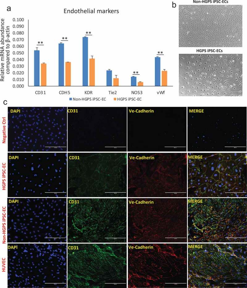
Characterization of iPSCs-derived ECs. (a) Real-time PCR for endothelial markers CD31, CDH5, KDR, Tie2, NOS3 and vWF in non-HGPS and HGPS FACS purified iPSC-derived EC. (b) Brightfield images of non-HGPS and HGPS purified iPSC-derived EC, showing different shape and size (see analysis in Figure 5(b–c)). (c) Immunofluorescence staining for endothelial markers CD31 and VE-Cadherin (CD144). Non-HGPS iPSC-ECs with no 1st antibodies added were used as negative control (ctrl); HUVECs were used as positive control.
Images show HGPS 167–1Q and non-HGPS 168–1P cells. N = 3 experiments per donor cells, Student t-test, **p ≤ 0.01.
