Figure 4.

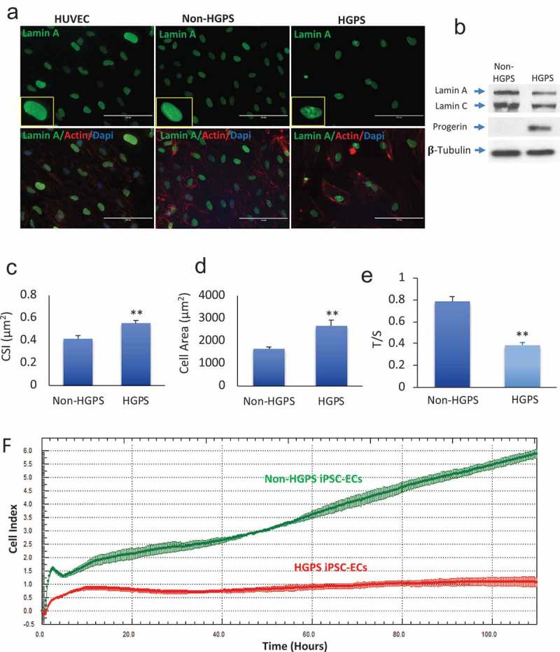
Senescence features in HGPS iPSC-derived ECs. (a) Immunofluorescence images showing the expression of lamin A in control HUVEC, non-HGPS and HGPS iPSC-EC. Nuclear dysmorphologies were observed in HGPS iPSC-ECs (right) compared to non-HGPS iPSC-ECs (left). (b) Western blotting for Lamin A/C and Progerin in Non-HGPS and HGPS iPSC-ECs. (c–d). HGPS iPSC-ECs were larger (cell area) and retain a rounded-shaped morphology [cell shape index (CSI)] compared to controls. (e) Bar graph of telomere length assessed by Monochrome Multiplex Quantitative PCR. HGPS iPSC-ECs have shortened telomeres compared to non-HGPS iPSC-ECs at the same passage, as shown by the reduced T/S ratios in HGPS iPSC-ECs. (f) Real-time cell analyzer profiles showed that HGPS iPSC-ECs (red curve) have a reduced cell index compared to non-HGPS iPSC-ECs (green curve).
Images show HGPS 167–1Q and non-HGPS 168–1P cells. N = 3 experiments per donor cells, Student t-test, **p ≤ 0.01.
