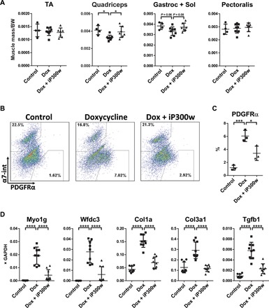
Fig. 3. Effect of iP300w on DUX4-induced alterations in vivo.

(A) Muscle mass of tibialis anterior (TA), quadriceps, gastrocnemius and soleus, and pectoralis, normalized to the body weight (BW) at day 12. iDUX4pA;HSA female mice were daily injected with dox (5 mg/kg, intraperitoneally) with or without iP300w (0.3 mg/kg) (n = 8). Wild-type (WT) siblings were used as a control (n = 4). (B) Representative FACS analyses for PDGFRα in pooled sample from quadriceps, gastrocnemius and soleus, pectoralis, and triceps at day 12. (C) Quantification of FACS analyses presented in (B). Data are presented as means ± SEM; *P < 0.05 and ***P < 0.001 by one-way ANOVA with Tukey’s post hoc test (n = 3). (D) RT-qPCR on RNA isolated from gastrocnemius at day 12. Note suppression of DUX4 target genes in the iP300w-treated group and reduction of expression of markers related to fibrosis. Data are presented as means ± SEM; ****P < 0.001 by one-way ANOVA with Tukey’s post hoc test. Results are presented as relative expression to glyceraldehyde-3-phosphate dehydrogenase (GAPDH) (n = 3).
