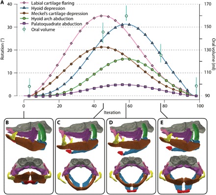
Fig. 3. Mouth opening and closing sequence.

(A) Reconstructed coordinated cranial cartilage motions and estimates of oral volume through mouth opening and closing cycle. (B) Closed mouth. (C) Mouth fully open with Meckel’s cartilage depressed. (D) Mouth closing with hyoid bar fully depressed. (E) Mouth closing with hyoid bar elevated. Vertical bars, extending from oral volume estimates, show maximum variance (table S2).
