
Fig. 2. Deficiency of mitochondrial PE promotes atrophy and respiratory failure.
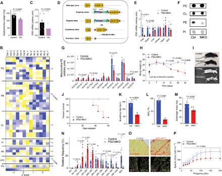
(A to C) Control (Ctrl; n = 6 or 7) and hindlimb-unloaded (HU; n = 7) C57BL6/J mice. (A) Gastrocnemius weight. (B) Skeletal muscle mitochondrial phospholipidome. (C) Skeletal muscle PSD mRNA. (D to P) Studies on PSD-MKO mice. (D) Generation of PSD-MKO mice. (E) PSD mRNA levels in multiple tissues (n = 5 to 6). (F) TLC analysis of mitochondrial phospholipids. (G) Muscle mitochondrial PE (n = 3 to 4). (H) Body weights after tamoxifen injection (n = 9 to 22). (I) Kyphosis in PSD-MKO mice. (J) Kaplan-Meier survival curve. (K and L) Breathing rate and peripheral capillary oxygen saturation (SpO2) 6 weeks after tamoxifen injection (n = 3). bpm, breaths per minute. (M to P) Diaphragm 4 weeks after tamoxifen injection. (M) Diaphragm weight (n = 4 to 6). (N) Distribution of fiber cross-sectional area (n = 9). (O) Fibrosis and fiber type. (P) Force-frequency curve (n = 4 to 6). Means ± SEM.