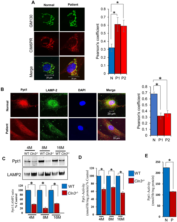
Figure 2: M6PR-mediated transport of PPT1 in normal and JNCL fibroblasts.
A. Confocal imaging showing colocalization of CI-M6PR fluorescence with that of Golgi marker, GM-130 [Control (n=30), Patient#1 (n=30) and Patient#2 (n=30)] *p<0.05. Note significantly increased colocalization suggesting impaired exit of CI-M6PR from the TGN in Cln3−/− mouse brain; B.Confocal imaging to show significantly reduced colocalization of Ppt1 and LAMP2 (lysosomal marker) immunofluorescence in cultured cells from JNCL fibroblasts compared with normal (N) fibroblasts. [Control (n=30), Patient#1 (n=30) and Patient#2 (n=30)] *p<0.05. Both Fig. 2A and 2B lower panel are representative image of fibroblast from patient 1. C. Western blot analysis of Ppt1-protein levels in brain lysosomes of 4-, 8-, and 16-mont h old WT and Cln3-mutant mice (n=4); *p<0.05; D. Lysosomal Ppt1-enzyme activity in 4-, 8-, and 16-month old WT and Cln3-mutant mice (n=6); *p<0.05; E. Ppt1 enzyme activity in cultured cells from normal (N) and JNCL patient (P). (n=3); *p<0.05.

