
Figure 2:
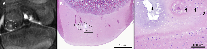
T2-weighted MRI of the left distal femur of miniature pig 2 (panel A) obtained 64 days after surgical interruption of the epiphyseal vascular supply to the abaxial aspect of the medial femoral condyle. The white circle marks an area of T2 hyperintensity consistent with epiphyseal cartilage necrosis within the medial femoral condyle. Photomicrograph (Panel B) depicts a coronal section through the medial femoral condyle. Black dashed line marks a discrete area of epiphyseal cartilage necrosis (OC-latens). Degenerate vessels within the area of epiphyseal cartilage necrosis are marked by black arrowheads. Area identified by the black rectangle is depicted in panel C at ten-times higher magnification. In panel C, a black asterisk marks an area of cystic matrix degeneration (an area where loss of tissue/matrix components results in clear spaces replacing the matrix) within an island of necrotic epiphyseal cartilage. Arrows indicate chondrocyte clones along the margin of the area of necrosis. Hematoxylin and eosin stain.