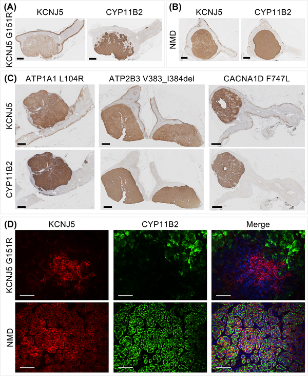
Figure 4: Heterogeneous immunostaining of KCNJ5 in APA according to genotype.
Immunohistochemical staining of KCNJ5 and CYP11B2 in an APA with a KCNJ5 mutation or in an APA with no mutation detected (NMD) showing decreased KCNJ5 immunostaining in the adenoma with a KCNJ5 mutation (Panel A, Panel B). APAs with ATP1A1, ATP2B3 or CACNA1D mutations displayed intense KCNJ5 immunostaining (Panel C). Double immunofluorescence staining of KCNJ5 and CYP11B2 in an APA with a KCNJ5 mutation compared with a NMD-APA (Panel D). KCNJ5 was intensely expressed in CYP11B2-negative cells in KCNJ5-mutated adenoma but markedly decreased KCNJ5 immunofluorescence was observed in CYP11B2-positive cells (Panel D, upper panel). In wild type APAs, KCNJ5 and CYP11B2 were co-localized to the same cells (Panel D, lower panel). DAPI staining (blue) was only included in the merged image. Panels A - C scale bar = 2 μm; panel D scale bar = 100 μm.

