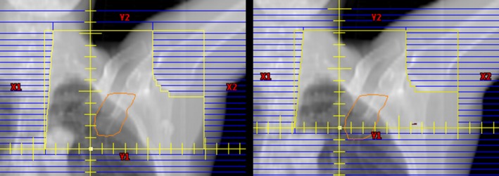
Figure 2.

Beam's eye view of supraclavicular fields for the original match line plan (left) and alternative match line plan (right) for the same patient. The contour of the level III axillary nodes are shown projected in orange. [Color figure can be viewed at wileyonlinelibrary.com]
