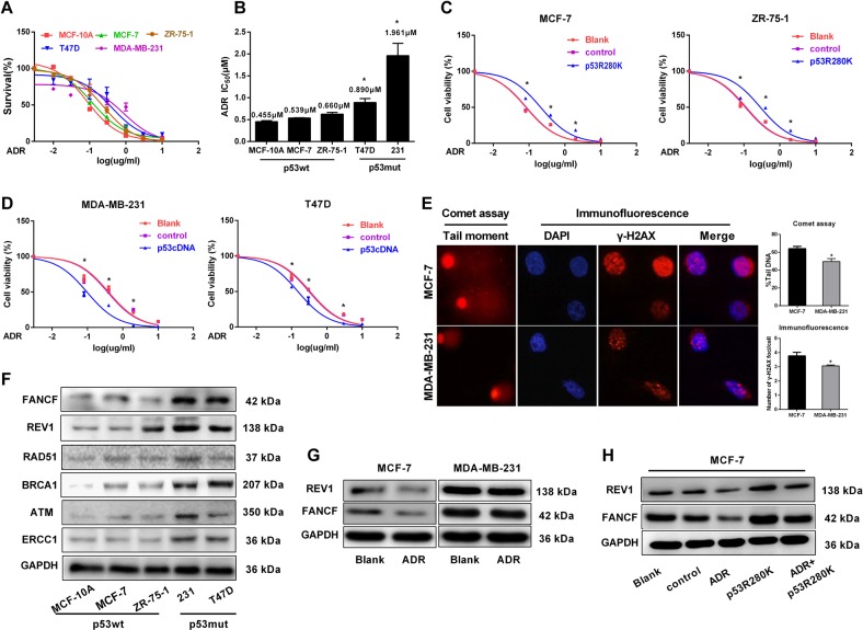
Fig. 1. p53 status confers ADR resistance through the upregulation of FANCF and REV1 expression.
a Determination of cell viability by Cell counting kit-8 (CCK-8) assays in the presence of various ADR concentrations with BrCa cells of known p53 status; mutP53 cells include MDA-MB-231 and T-47D, and wtp53 cells include MCF-10A, MCF-7, and ZR-75-1. b The IC50 value of BrCa cells with different p53 status. The IC50 value was calculated using Graphpad 7.0 software. *p < 0.05 vs. p53wt BrCa cells. c MCF-7 and ZR-75-1 cells expressing an empty vector (control) or transduced with mutp53 protein (p53R280K) and untreated MCF-7 and ZR-75-1 cells (blank) were treated with various ADR concentrations. Cell viability was assessed by CCK-8 assays. *p < 0.05 vs. control group. d MDA-MB-231 and T47D cells expressing an empty vector (control) or transduced with wtp53 protein (p53cDNA) and untreated MDA-MB-231 and T47D cells (blank) were treated with various ADR concentrations. Cell viability was assessed by CCK-8 assays. *p < 0.05 vs. control group. e After MCF-7 and MDA-MB-231 cells were treated with ADR (0.5 µM) for 48 h, we conducted an analysis of DNA damage by comet assays and γ-H2AX foci formation. The images were captured using a fluorescence microscope at ×400 magnification. Comet tails represent fragmented DNA leaked from the nucleus. Tail intensity (% Tail DNA), defined as the percentage of Comet tails migrated from the head of the comet to the tail, was applied as a measure of DNA damage;48 50 comet tails were analyzed for each sample. Quantification results are shown to the right. *p < 0.05 vs. MCF-7 group. For each sample, 50 nuclei were analyzed. f Western blot analysis of DNA repair-related proteins in BrCa cell with different p53 status. g MCF-7 and MDA-MB-231 cells were untreated and treated with ADR (0.5 µM) for 48 h. The cells were lysed for Western blotting and probed with the indicated antibodies. h Immunoblot analysis of MCF-7 cells transfected with an empty vector (control) or the p53R280K plasmid and then treated with ADR (0.5 µM) for 48 h. Data represent mean ± SD of three independent experiments. (*p < 0.05, **p < 0.01, ***p < 0.001)

