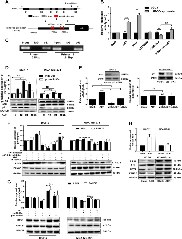
Fig. 5. p53 regulates the expression of FANCF and REV1 via miR-30c in BrCa.
a Schematic representation of NFYC (miR-30c host gene) and putative p53 binding sites in intron 5 of NFYC. b HEK-293T cells were treated with pcDNA3.1-p53 (wtp53), pcDNA3.1-p53R280K (mutp53), ADR (0.5 µM), pifithrin-α (10 µM), pifithrin-α + ADR or the miR-30c promoter constructs (in the pGL3 vector). Relative luciferase activity was assayed. **p < 0.01 vs. PGL3 vector, ##p < 0.01 vs. pifithrin-α group. c ChIP assay showing endogenous p53 bound to the miR-30c promoter in the p53 binding site region in MCF-7 cells. d MCF-7 and MDA-MB-231 cells were treated with ADR (0.5 µM) for 12, 24, and 48 h. The expression of mature miR-30c and pri-miR-30c was evaluated by qRT-PCR. The protein expression of p-p53, p53, and p21 was examined by Western blotting. *p < 0.05, **p < 0.01 vs. 0 h group. e MCF-7 and MDA-MB-231 cells were treated with control, p53shRNA, ADR, or p53shRNA + ADR for 48 h. qRT-PCR showed that the miR-30c expression changes. The protein expression of p53 was examined by Western blotting. f MCF-7 and MDA-MB-231 cells were transfected with NC inhibitor (20 nM) or miR-30c inhibitor (20 nM) and were then treated with ADR and p53cDNA. The expression of FANCF and REV1 was analyzed by Western blotting. MCF-7 cells: *p < 0.05, **p < 0.01 vs. Blank group; #p < 0.05 vs. ADR group, ##p < 0.01, vs. p53cDNA group. MDA-MD-231 cells: *p < 0.05 vs. Blank group; #p < 0.05 vs. p53cDNA group. g FANCF and REV1 expression in MCF-7 and MDA-MB-231 cells transfected with p53 shRNA, miR-30c mimic (miR-30c) or the negative control (NC). The expression of FANCF and REV1 was analyzed by Western blotting. **p < 0.01, ***p < 0.001 vs. Blank group; #p < 0.05 vs. Blank group. h qRT-PCR analysis of miR-30c expression in ADR-treated MCF-7 and MDA-MB-231 xenografts. Western blot analysis of p-p53, p53, FANCF, and REV1 expression in ADR-treated MCF-7 and MDA-MB-231 xenografts. **p < 0.01, ***p < 0.001 vs. Blank group. NS indicates no significant difference. Data represent the mean ± SD (n = 3, each group)

