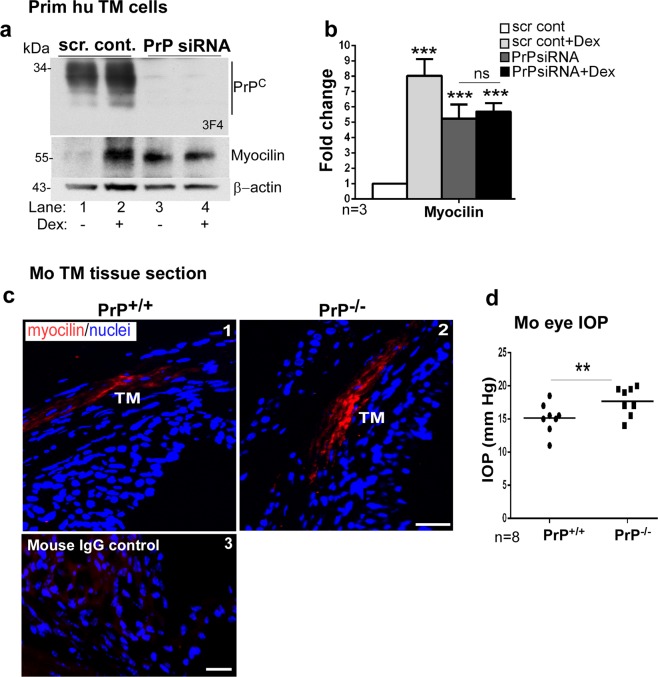
Figure 7.
Myocilin is upregulated by downregulation of PrPC. (a) PrPC was silenced in primary human TM cells and lysates were processed as above. Probing for PrPC shows the expected glycoforms in controls, and minimal reaction in samples treated with PrP-siRNA (lanes 1 & 2 vs. 3 & 4). Re-probing for myocilin shows significant upregulation in the absence of PrPC (lane 1 vs 3). Exposure of control and experimental cells to dexamethasone (Dex) shows upregulation of myocilin as expected (lanes 1 & 2). However, no additive effect of dexamethasone is noted in the absence of PrPC (lanes 3 & 4). (b) Quantification by densitometry after normalization with β-actin shows 8-fold upregulation of myocilin by dexamethasone, and ~5.3-fold upregulation in the absence of PrPC regardless of dexamethasone. Values are mean ± SEM of the indicated n. **p < 0.01; ns: not significant. Full-length blots are included in the Supplementary Fig. S2. (c) Immunoreaction of anterior segment of PrP+/+ and PrP−/− mice for myocilin shows upregulation in the TM of PrP−/− sections relative to controls (panels 1 & 2). No reaction was detected in a serial section reacted with mouse IgG followed by Alexa 546-conjugated secondary antibody (panel 3). Scale bar: 25 µm. (d) Measurement of IOP shows significant upregulation in PrP−/− eyes relative to PrP+/+ controls (n = 8). Values are mean ± SEM of the indicated n. **p < 0.01.

