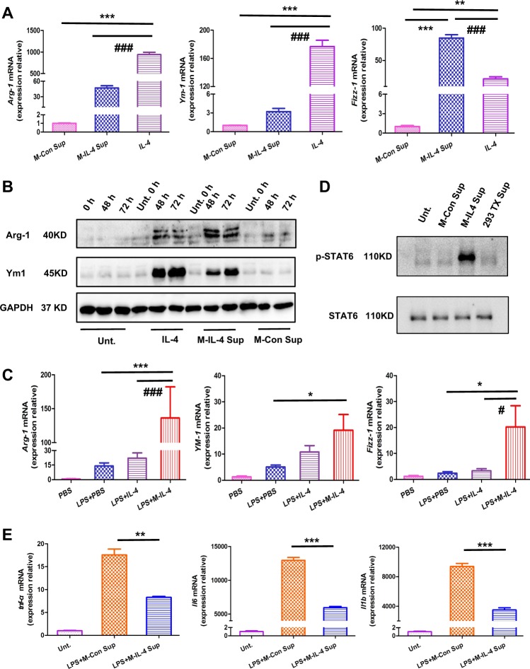
Fig. 2. M2-driving ability and the anti-inflammatory effect of murine macrophages secreting IL-4 (M-IL-4).
a Bone marrow-derived macrophages (BMDMs) were treated with IL-4, M-IL-4 supernatant, or M-Con supernatant for 48 h. Cells were collected and the mRNA level (Arg-1, Ym-1, and Fizz-1) was determined by qPCR. b BMDMs were treated with IL-4, M-IL-4 supernatant, or M-Con supernatant and the cells collected at the indicated time. The protein levels of M2 markers (Arg-1, and Ym-1) were determined by western blotting (WB). c Mice were treated with LPS (15 mg/kg) and after 0.5 h, PBS, IL-4 (10 ng/g), or M-IL-4 (2.5 × 10⁵ cells/mouse) was intratracheally transplanted. The lungs were isolated after 48 h (n = 6 for each group), and the expression of M2 markers (Arg-1, Ym-1, and Fizz-1) was determined by qPCR. d IL-4 and M-IL-4 supernatant activated the STAT6 signaling pathway. BMDMs were treated with M-IL-4 supernatant, M-Con supernatant, 293TX supernatant, or no supernatant, and after 24 h, cells were collected for WB. e BMDMs were treated with/without LPS and after 0.5 h, M-IL-4 supernatant or M-Con supernatant was added for 6 h, followed by the collection of cells. The mRNA expression of M1 markers (tnf-α, Il6, and Il1b) was determined. *P < 0.05, **P < 0.01; #P < 0.05, ###P < 0.001, the error bars represent ± SD

