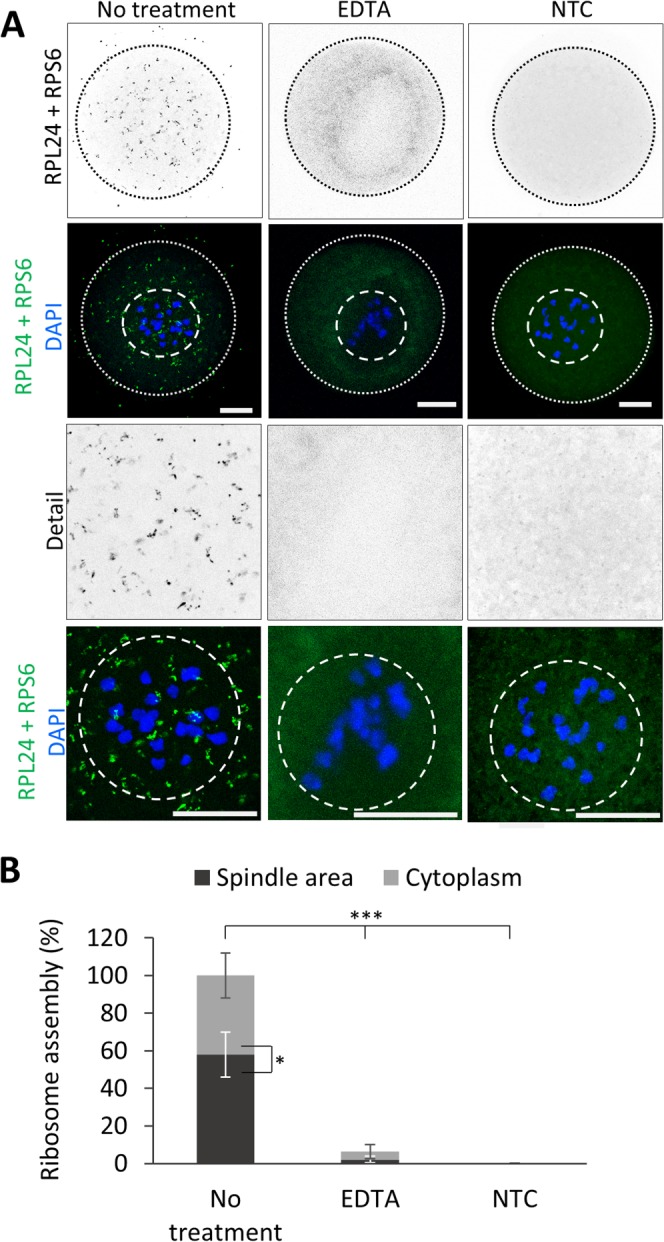
Figure 4.

Spindle area contains assembled ribosomes. (A) Proximity ligation assay (PLA) detecting ribosome assembly using RPL24 and RPS6 markers (grey and green dots). EDTA was used for ribosome disruption. As a negative control (NTC) RPS6 antibody was omitted. The cortex of the oocytes is depicted by a black or white dotted line; dashed line shows spindle area. Representative images of three independent experiments (n ≥ 15); scale bar = 20 μm. (B) Quantification of ribosome assembly events in NEBD oocytes. Mean; error bars are SD; Student’s t-test, *P < 0.05; ***P < 0.001.
