Figure 5.
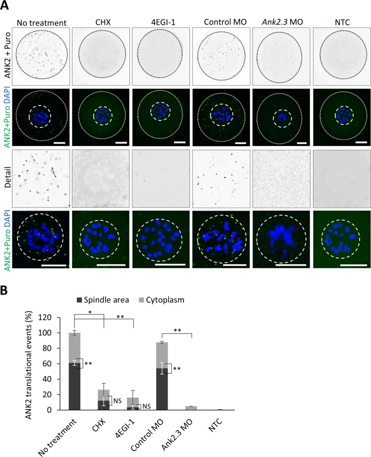
ANK2 is translated at the newly forming spindle through cap-dependent translation. (A) Ribopuromycilation combined with Proximity ligation assay (Puro-PLA) were performed on NEBD stage oocytes in the absence or presence of cycloheximide (CHX) and eIF4E/eIF4G1 interaction inhibitor (4EGI-1). Microinjection of morpholinos (MO) was used to specifically suppress Ank2.3 mRNA translation in the oocyte. ANK2 translational events in grey and green scales, DNA in blue. The cortex of the cell is depicted by a black or white dotted line; dashed line shows spindle area. Representative images from at least three independent experiments (n ≥ 15), scale bar = 20 μm. As a negative control (NTC) ANK2 antibody was omitted. (B) Quantification of ANK2 translational events at the spindle area (black) and cytoplasm (grey). Mean, error bars are SD; Student’s t-test; *P < 0.05; **P < 0.01; ***P < 0.001; NS non-significant. See also Fig. 6D and Suppl. Figs 3 and 5.

