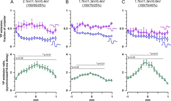
Figure 5.
Stronger bipolar cancellation rendered by multiphasic nsEP improves stimulation targeting. Cell electropermeabilization assay and stimulation conditions are the same as described in Fig. 4. For panels (A–C), the shapes of stimulating pulses and local waveforms arising from their superposition are provided in Fig. 3, (B–D), respectively. The amplitude of each phase (kV) and their ratio (%) for a triphasic nsEP applied to electrode 2 are also given at the top of each panel; biphasic pulses applied to electrode 3 are the same but omit the first phase. Top panels show the degree of bipolar cancellation when additional phase(s) are added to a unipolar pulse. The data plots for tri- and biphasic pulses are identified by respective symbols. YP emission caused by stimulation with tri- and biphasic pulses is plotted as a fraction of the effect of the respective unipolar pulse at the same location. Abscissa is the distance from the center of the gap between electrodes 2 and 3 along a line connecting them (Fig. 2A). See Fig. S2 for YP emission values without normalization to unipolar pulse data. Bipolar cancellation is significant at p < 0.05 or better for all datapoints (one sample t-test, for the ratio being different from 1). Bottom panels: The enhancement of effect remotely from the electrodes when the tri- and bi-polar pulses are delivered synchronously (with one phase shift) to electrodes 2 and 3 respectively. For comparison, the same pulses are delivered with a 10 ms delay, to prevent their overlap and opposite polarity compensation (same as in Fig. 4F). See Fig. S2 for actual values of the effects of these pulse delivery protocols. Mean ± S.E., n = 5–6. *p < 0.5, **p < 0.01, one sample t-test.

