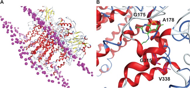
FIG 5.
Molecular modeling of CWHM-728 in the predicted structure of M. tuberculosis QcrB. (A) QcrB homology model embedded in the lipid bilayer, with the lipid head group phosphorus being shown as magenta spheres. (B) QcrB binding site shown with CHWM-728 docked (green). The residues mutated in resistant mutants are highlighted in magenta.

