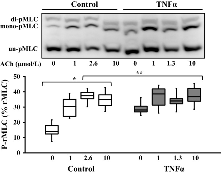
Figure 6.

Representative western blots in which phosphorylated (mono‐ and di‐ p‐rMLC) and unphosphorylated (un‐p‐rMLC) rMLC were separated using a modified Phos‐tag™ gels. In control ASM, ACh stimulation increased rMLC phosphorylation (p‐rMLC relative to total rMLC) up to the EC50 concentration (2.6 μmol/L) but with no change at 10 μmol/L. Exposure to TNFα (100 ng/mL, 24 h) significantly increased the basal level of rMLC phosphorylation but blunted the ACh‐dependent increase in pMLC. The ratio of p‐rMLC to total rMLC increased with 1 and 2.6 μmol/L ACh stimulation in control ASM strips and phosphorylation of rMLC did not increase at the 10 μmol/L ACh stimulation. In the TNFα‐treated group, basal p‐rMLC was increased in unstimulated ASM strips and there was no significant difference at the other doses of ACh stimulation. Values are medians – IQR. *Significant difference (P < 0.05) compared to unstimulated ASM strip within the control group (n = 6 animals). **Significant interaction between ACh concentration dependence and treatment group. rMLC, regulatory myosin light chain; ASM, airway smooth muscle; ACh, acetylcholine; TNFα, tumor necrosis factor alpha.
