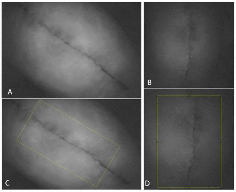
Figure 1. A baseline image is shown in (A), followed by an image captured immediately after the ice was removed from the anterior (incisional) knee (B). The ImageJ capture points are shown in (C) and (D) for the respective photos.
The indocyanine green dye within the blood vessels shows up as white on the images, while dark areas are devoid of blood flow.

