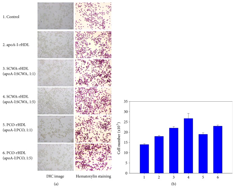
Figure 5.
Enhancement of cell growth by rHDL containing SCWA and PCO in brain glial (BV-2) cells. Cell nucleus was visualized by hematoxylin staining in the presence of SCWA or PCO in rHDL. Photo 1, control; photo 2, apoA-I- rHDL; photo 3, SCWA-rHDL (1:1); photo 4, SCWA-rHDL (1:5); photo 5, PCO-rHDL (1:1); photo 6, PCO-rHDL (1:5).

